


Breast biopsy


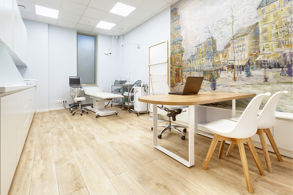

The indication for core needle biopsy or mammotome-assisted breast biopsy is a focal lesion of interest detected in imaging tests (breast ultrasound, mammography) or by palpation. The aim of both procedures is to establish a histopathological diagnosis based on the biopsy samples.
Core needle biopsy, although less invasive, is used to take smaller tissue samples from the breast and requires separate insertion of the needle for each sample taken. Mammotome-assisted biopsy uses a vacuum system to obtain a larger sample of tissue material in one needle insertion.
Both procedures are performed on an outpatient basis, under local anesthesia. The patient can go home immediately after the biopsy.
Core needle biopsy
The doctor qualifies the patient for the procedure and then performs the biopsy with the patient supine or slightly on their side. The biopsy site is disinfected before the procedure. The lesion is located under ultrasound guidance and 2% lignocaine solution is injected for local anesthesia. Then an approximately 1.5 to 3 mm diameter needle is inserted into the target site and the trigger mechanism is activated to collect a tissue sample. This is repeated several times to obtain the appropriate amount of material. The collected material is sent for histopathological evaluation, and the biopsy site is cleaned, disinfected, and wound dressing is applied. Core needle biopsy of the breast takes about 15 to 20 minutes to complete.
Mammotome-assisted biopsy
The biopsy site is disinfected before the procedure just like for core needle biopsy. The lesion is located under ultrasound guidance and local anesthesia is administered. Then an approximately 2 to 3 mm diameter needle is inserted into the target site and the vacuum mechanism is activated to collect a tissue sample through a special groove. The collected material is sent for histopathological evaluation, and the biopsy site is cleaned, disinfected, and wound dressing is applied. Mammotome-assisted biopsy of the breast takes about 15 to 20 minutes to complete.
Consult our specialists: